MECHANISMS OF CELL ATTACHMENT
Introduction: Fibrous elements of the cytoskeleton, including microtubules and filaments, help maintain cell shape. Cell shape is determined by the interaction of these elements with the surface to which the cell is attached.
Importance: Because of the difficulty in visualizing the underside of cells, light reflection through a cell and glass slide can be used to describe the attachment of cells to surfaces. By measuring how the refractive index of a cell changes, we can determine how a cell's density and cytoskeleton changes when it attaches to a surface.
Question: How do the density and cytoskeleton of a cell change when it attaches to a surface?
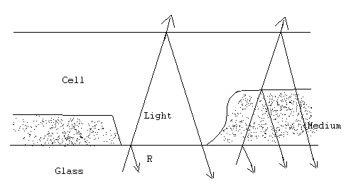
Method: For a cell in a surrounding medium attached to a glass slide, light is reflected onto particular regions of the cell. If the cell is directly attached to the glass, then the intensity of light reflected at this interface can be measured. If, instead, the surrounding medium separates the cell from the glass, then the intensity of light reflected is determined by the glass/medium interface. The refractive index of the glass, cell, and medium determines how much light is reflected at each of these interfaces.

The relative intensity (R) of reflected light at the interface of the glass and cell is given by

where rg and rc are the refractive indices of the glass and cell respectively. The refractive index of the glass used here is approximately 1.515 and is generally greater than the refractive index of the cell. As cell density increases, the refractive index of the cell increases. Notice that as rc increases, the denominator increases, but the numerator decreases. Therefore this is a decreasing function, and the relative intensity of reflected light (R) decreases as the refractive index (rc), or cell density, increases.
By rearranging this equation, the refractive index (rc) of a particular region of the cell can be calculated by measuring the intensity of reflected light at that region:

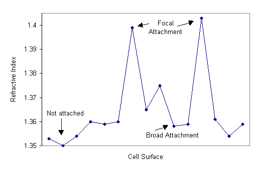
The refractive index can be calculated for different regions along a cell surface to compare the density of the cell at regions where it is focally, broadly, and not attached to a surface.

Interpretation: In regions where cells were attached to glass by a single focal point, refractive indices were higher than regions of the cell that were broadly attached to the glass or not attached to the glass at all. The higher refractive indicates regions of focal contact reflect an increase in the density of the cytoplasm at regions of attachment.
Conclusion: In regions of a cell attached to a substrate, the density of that local region of the cell increases. This results in a decrease in the amount of light reflected. The increased density at attachment sites results from microfilaments in the cytoskeleton contracting to support the cell.
Source: Bereiter-Hahn, J., C. H. Fox, and B. Thorell, 1979, Quantitative reflection contrast microscopy of living cells, J. Cell Biology 82:767-779
Izzard, C. S. and L. R. Lochner. 1976. Cell-to-substrate contacts in living fibroblasts: An interference reflexion study with an evaluation of the technique. Journal of Cell Science 21:129-159.
Copyright 1999 M. Beals, L. Gross, S. Harrell