QUANTITATIVE CYTOLOGICAL CHARACTERISTICS: CELLS UNDER STRESS
Introduction: In addition to the cell membrane, cells contain a great deal of internal membranes that form and support organelles. Mitochondria, for example, are composed of an outer membrane and a folded, inner membrane. The volume of a cell or organelle and the surface area of the corresponding membrane surface area can greatly influence the rates of diffusion of ions and solutes into the cell or organelle. Consequently, properties of cells might change under different conditions in order to improve rates of diffusion.
Importance: Cytological characteristics of a cell can vary from cell to cell and frequently depend on properties of the cell's environment. We might expect cytological characteristics to change when a cell needs to increase or decrease rates of diffusion. For example, plant cells may be stressed under photorespiratory conditions (high temperature, low CO2, high O2). Photorespiration interferes with photosynthesis and inhibits ATP formation. We can use quantitative cytological techniques to gain information about cells under stressful environmental conditions.
Question: How do quantitative characteristics of the cell change when a cell is under stress?
Variables:
|
V |
volume density (% cell occupied by structure) |
|
O |
number of grid points on organelle |
|
N |
number of organelles in cell analyzed |
|
C |
number of grid points on cell |
|
S |
surface to volume ratio |
|
M |
number of grid points on membrane |
|
D |
diameter of cell wall |
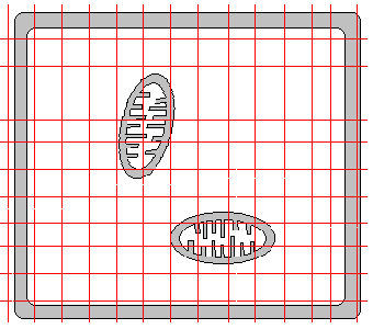
Methods: Stereology techniques can be used to gain information from 2 dimensional images of cells. A grid of lines is placed over a slide of a cell, and points of interest in the cell that intersect the grid lines are counted.
Volume density (V) for a particular organelle can be calculated as

where O is the number of grid points falling on a total of N organelles and C is the number of grid points falling on the entire cell.
Surface to volume ratio (S) can be calculated for chloroplast thylakoids or mitochondrial cristae as
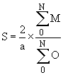
where M is the number of grid points falling on the folded inner membrane of these structures (a total of N structures in the cell are analyzed), O is the number of grid points falling on the entire chloroplast or mitochondria (N of them), and a is a correction factor related to the size of the grid.
Surface to volume ratios can similarly be calculated for the cell membrane and cell wall using
![]()
where M is the number of grid points intersecting the membrane or cell wall and D is the diameter of the cell wall.
The fold index (F) of the plasma membrane can be determined as

where Sm and Sw are the surface to volume ratios of the plasma membrane and cell wall.
We can compare quantitative cell characteristics for a plant under stressful conditions, Elodea, with a typical C3 plant, the sunflower.
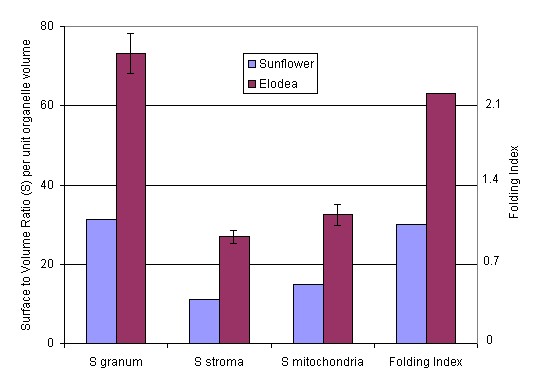
Interpretation: In Elodea, an aquatic monocot, the surface area to volume ratio for mitochondria was found to be quite high under photorespiratory conditions (Smitochondria). This might reflect the need for high ATP production. Surface to volume ratios for chloroplasts (Sgranum and Sstroma) were also much higher than a typical plant under unstressed conditions. Additionally, a high folding index (F) indicated a great deal of infolding in the plasma membrane. Presumably, the increased surface area would allow for greater transport of ions into the cell.
Conclusions: Properties of cells, including surface area to volume ratios, can give us a great deal of information about how cells respond to stressful conditions. Quantifying these properties using stereology techniques allows for more objective comparisons between cells from different plants or under different conditions.
Additional Questions:
1. Compare Elodea and sunflower to maize, a C4 plant. For maize, Sgranum and Sstroma values are 67.3 and 29.4, respectively. Knowing the properties of C4 plants, would you expect Elodea (under conditions of low CO2) to be more similar to sunflower or maize, and why?
Source: W. R. Fagerberg, T. T. Eighmy, and L. S. Jahnke. 1991. Studies of Elodea nuttallii grown under photorespiratory conditions. III. Quantitative cytological characteristics. Plant, Cell, and Environment 14, 167-173
Weibel, E. R. 1979. Stereological Methods Volume 1: Practical Methods for Biological Morhpometry. Academic Press, NY, NY.
Copyright 1999 M. Beals, L. Gross, S. Harrell